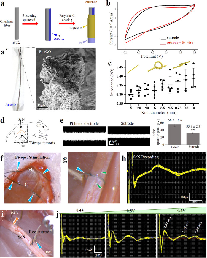
Fig. 1. Platinized graphene fibers serve as suture and wrapping electrodes.
a Coating steps of extruded Pt-rGO electrodes. a’ Photograph and SEM. b Cyclic voltammetry of Pt-rGO fibers before and after soldering to a Pt wire. c Graphene fiber knotting does not significantly alter the electrode impedance. Individual impedance measurements and their mean and standard deviation are represented in the graphic during different knot diameters in the sutrode (n = 5 sutrodes). d Illustration of the ScN and biceps femoris muscle. e Reduced base noise measurements of the sutrode compared to Pt hook electrodes in saline, graphics in the right were obtained from n = 5 independent experiments **p < 0.01 (values represented as mean ± SD). f Sutrode placed on biceps (blue arrows) evoked effective muscle contraction (Supplementary Video 1). g Sutrode wrapped snugly on the ScN (blue arrow), without blood flow occlusion of superficial vasculature (green arrows) used to h record the evoked CNAP. i Stimulation of the tibial nerve fascicle with hook electrodes and recording with the sutrode (blue arrow), allowed the recording of j graded evoked CNAPs and the detection of B and C fibers. Pt-rGO reduced liquid crystalline graphene oxide, s straight sutrode, ScN sciatic nerve, SEM scanning electron microscopy, CNAP compound nerve action potentials.

