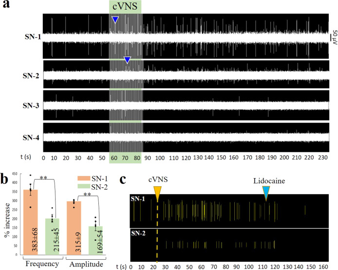
Fig. 6. Differential neural activity in SN branches in response to cVNS.
a cVNS with 0.5 V, 30 s pulses evoked an immediate increase in SN-1 and SN-2 (blue arrowheads) and less in SN-3. No activity was evoked in SN-4. b Comparison between SN-1 and SN-2 spike frequency and amplitude signal increase, values were calculated at 60 s of stimulation and presented as mean ± SD for n = 5 animals; **p < 0.01. c Lidocaine (blue arrow) blocked the neuronal activity evoked by cVNS (yellow arrow), confirming its neuronal nature. SN splenic nerve, cVNS cervical vagus nerve stimulation, SD standard deviation.

