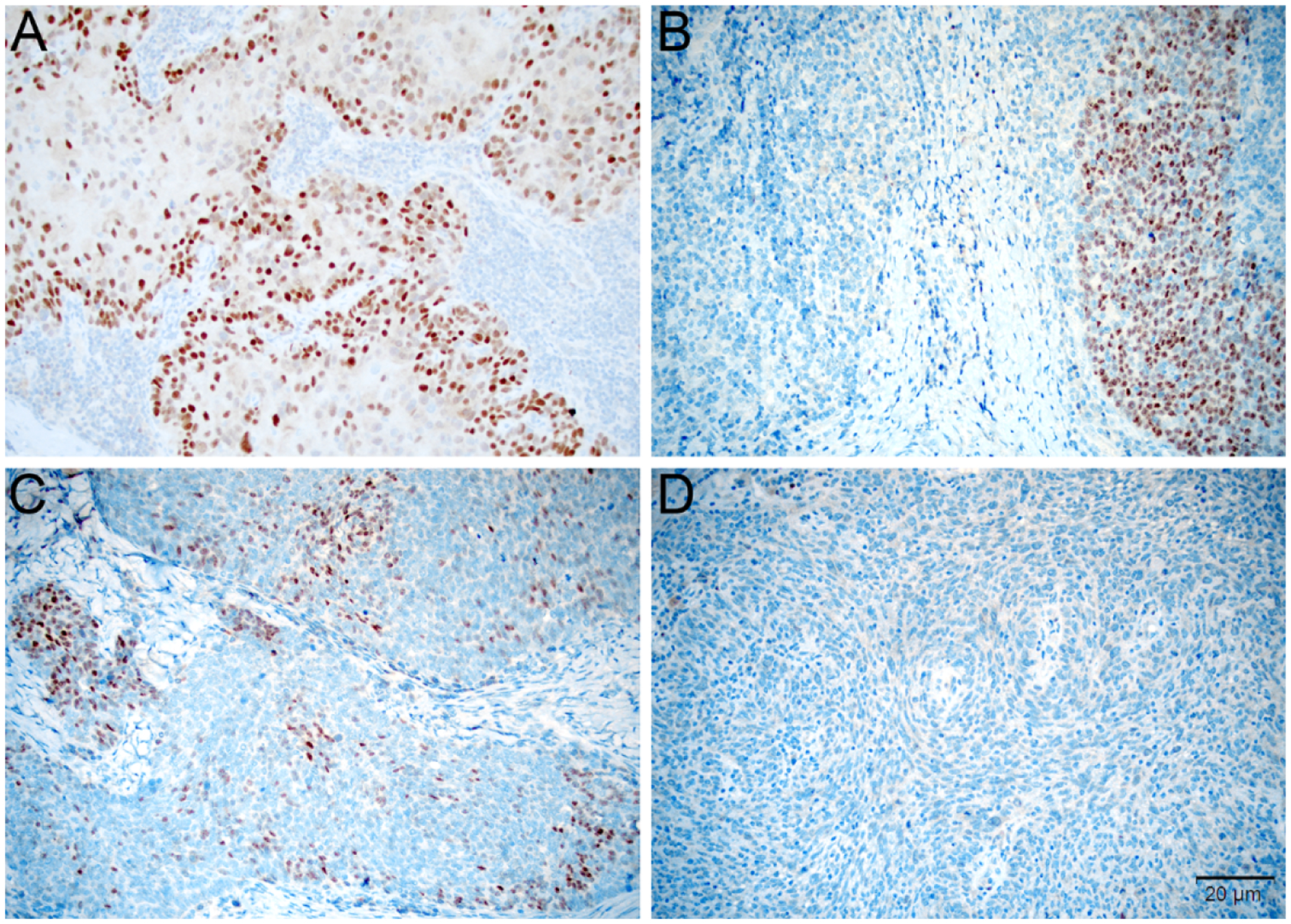
Figure 1:

Tumors were scored for ERα based on the percentage of positive tumor cells, intensity of staining, and pattern of staining. Tumors were considered diffusely positive if most cells were uniformly positive throughout the tumor (A, 200x), to have block-like staining if discrete positive areas alternated with negative areas (B, 200x), and patchy if a subset of cells were stained at a similar level throughout the tumor (C, 200x). Based on the thresholds developed in breast carcinoma, staining was considered negative if <1% of cells showed reactivity (D, 200x).
