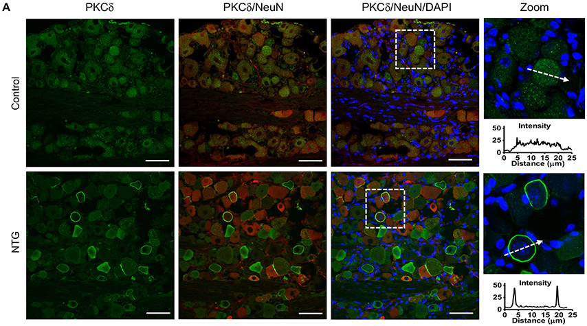
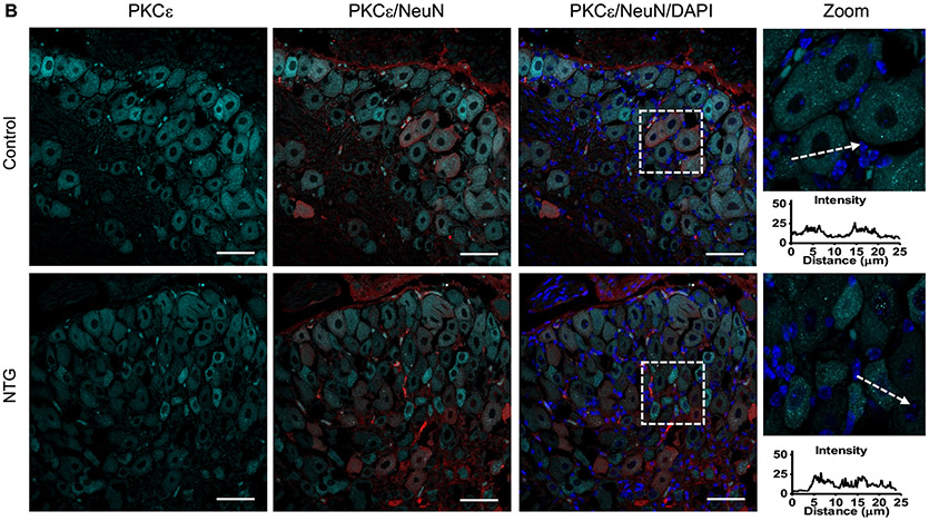
Figure 5.
Activation of PKCδ in the trigeminal ganglion (TG) neurons in chronic NTG-treated mice. Immunohistochemistry analysis showed plasma membrane translocation of PKCδ (A), but not PKCε (B), on Day 7 in TG of chronic NTG-treated mice. The fluorescent intensity of PKC isoform across representative cells (indicated by dashed arrows) is shown. Green, PKCδ; Cyan, PKCε; Red, NeuN; Blue, DAPI. Scale bars: 50 μm. n = 15 slices from 3 mice.


