
Figure 2. Effects of serum exosomes on stroke outcome are age-dependent.
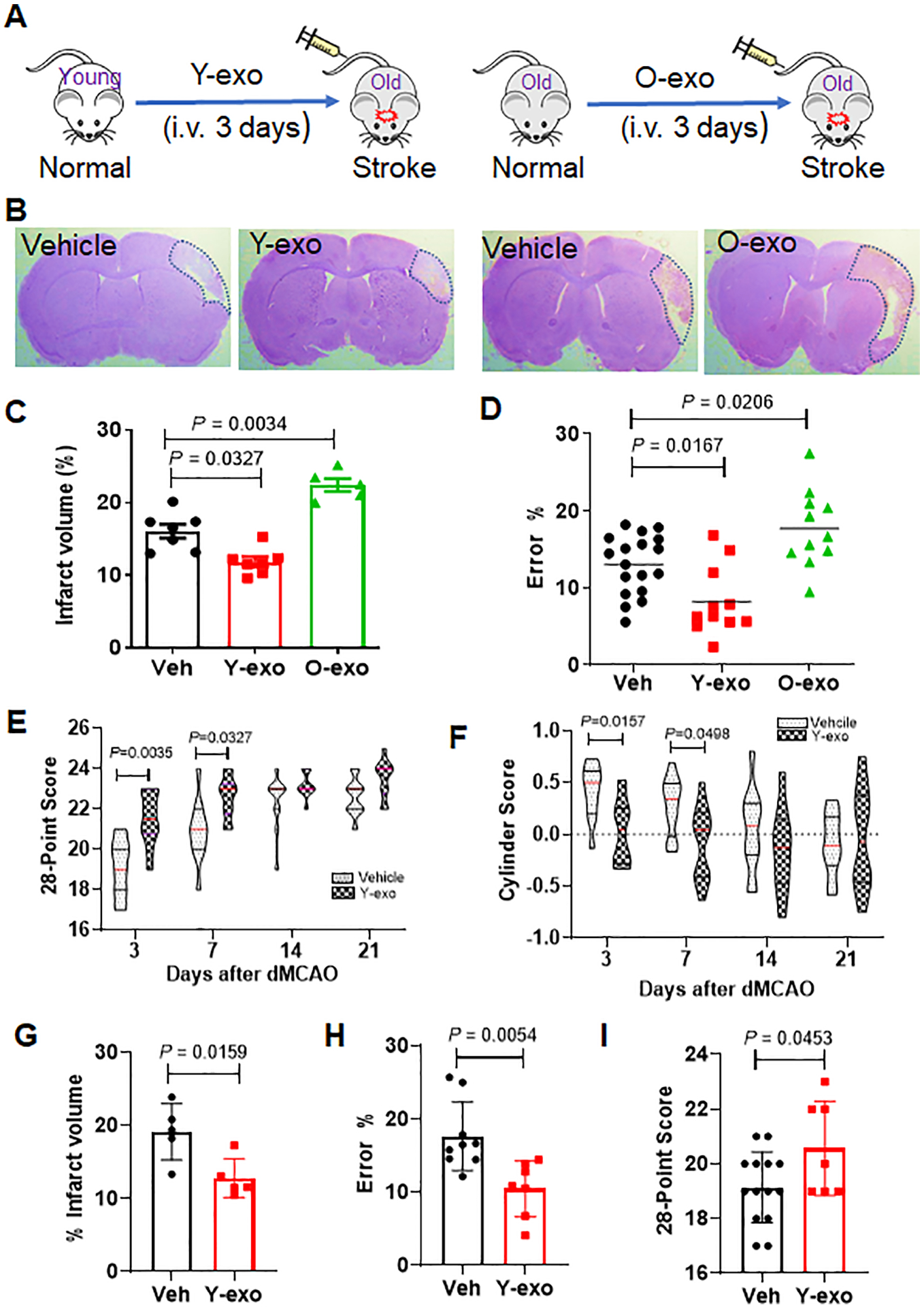
A. Schematic illustration of intravenous injection of Y-exo (right panel) and O-exo (left panel) into aged ischemic rats 3 h after ischemic stroke, twice per day for 3 days. B. Representative images of CV-stained coronal brain sections of vehicle–, Y-exo– and O-exo–treated rats 72 h after focal ischemia. C. Quantitative analysis of infarct volume in vehicle–, Y-exo– or O-exo– treated rats 72 h after ischemic stroke. The sample size was N = 7 in the Veh and Y-exo groups, and N = 5 in the O-exo group. The P values were assessed by a one-way ANOVA followed by a Tukey’s multiple comparisons test. D. Sensorimotor deficits of aged ischemic rats were assessed by the ladder rung walking test 72 h after administration of vehicle (N = 18), Y-exo (N = 11), or O-exo (N = 11). P values were shown and assessed by one-way ANOVA followed by Tukey’s multiple comparisons test. E-F. The long-term effect of Y-exo treatment on sensorimotor function assessed by 28-point neuroscore tests (E) and Cylinder test (F) 3, 7, 14 and 21 d after dMCAO (N = 11 per group). The P values were assessed by a 2-way repeated measures ANOVA with a Bonferroni post-hoc test. G. Delayed treatment (6 h after onset of ischemia) of Y-exo significantly decreased infarct volume in aged rats 72 h after dMCAO (N = 5 per group). The P values assessed by a Mann-Whitney test. H and I. The effect of delayed treatment of Y-exo on sensorimotor function assessed by the ladder rung walking test (H) and 28-point neuroscore tests (I). The sample size was N = 14 in the Veh group and N = 7 in the Y-exo group. Data are presented as the mean ± SEM. The P values were assessed by an unpaired Student’s t-test. Y-exo, serum exosomes from young rats; O-exo, serum exosomes from aged rats. Dotted lines in B indicate the border of infarct core.