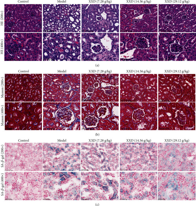
Figure 6.

Effect of XXD on renal histopathology of the kidneys. (a) Hematoxylin and eosin (HE) and (b) Masson staining show changes in the control and model mice with XXD treatment. Bars represent 100 μm and 50 μm. (c) SA-β-gal staining of kidney tissue in AKI mice. Semiquantitative analysis of SA-β-gal staining showed a significant decrease of SA-β-gal activity in the XXD-treated kidneys as compared with the model mice.
