Fig. 1.
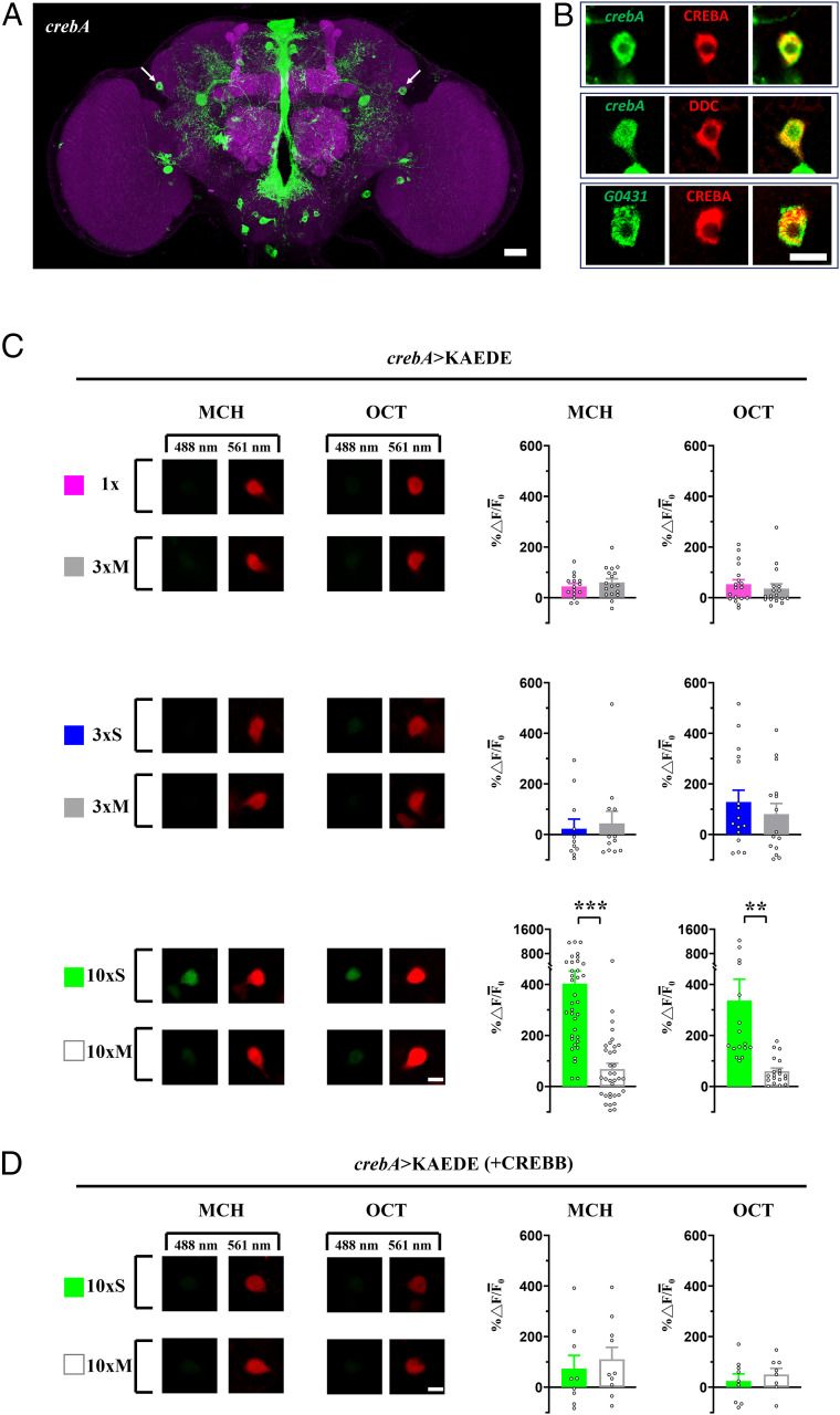
crebA expression in DAL neurons was enhanced after 10×S training and repressed by CREBB. (A) CREBA in dissected brains (green), counterstained with anti-DLG immunostaining (magenta). Arrows = DAL neurons. (Scale bar, 20 µm.) (B) Single optical slices of DAL neuron. crebA expression (green) colocalized with CREBA antibody (red) (Top) and with DDC antibody (red) (Middle) and G0431-Gal4 expression (green) colocalized with CREBA antibody (red) (Bottom). (Scale bar, 10 µm.) (C) crebA promoter activity after 1×, 3×M, and 3×S training with both MCH and OCT as reported by de novo KAEDE fluorescent protein synthesis (Left), estimated by the ratio of new (green, 488 nm) and preexisting (red, 561 nm) proteins (% ∆ F/¯F0) (Right). For each brain, single DAL neuron optical slices were imaged under identical conditions. Training effects of 1× compared with 3×M (Top) and effects of 3×S compared with 3×M (Middle) were not significant. The crebA promoter was activated after 10×S but not 10×M (Bottom). (Scale bar, 10 µm.) (D) crebA activity was repressed by crebB overexpression (+CREBB) after 10×S as compared with 10×M training. (Scale bar, 10 µm.) Bars represent mean ± SE, n ≥ 8/bar. **P < 0.01; ***P < 0.001.

