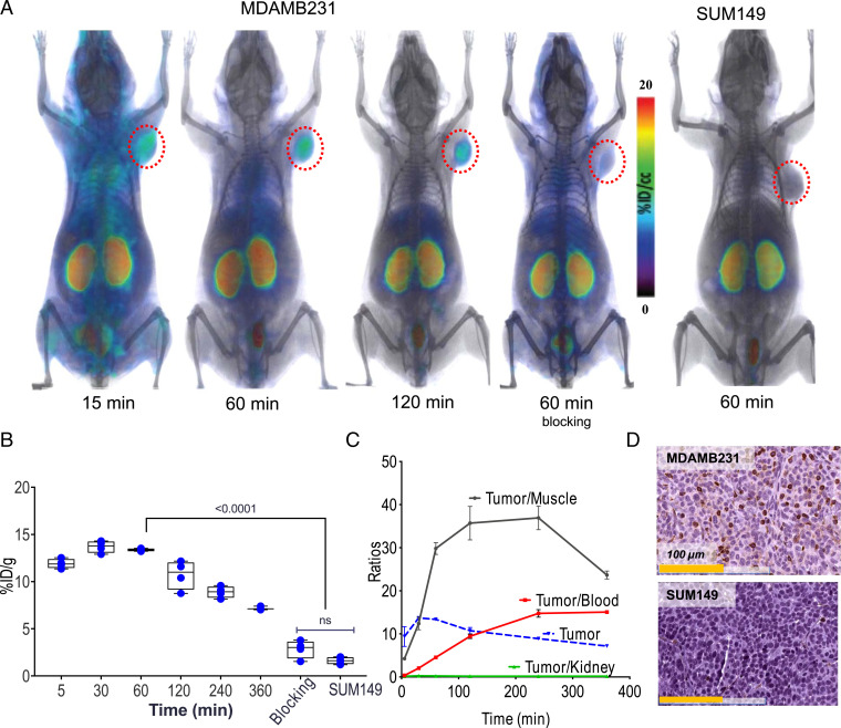
Fig. 3.
In vivo kinetics of [18F]DK222 in mice bearing TNBC xenografts. NSG mice with human tumor xenografts were injected with 200 μCi (7.4 MBq) [18F]DK222, and PET-CT images were acquired at different time points. Blocking-dose mice received 50 mg/kg of DK221 30 min prior to radiotracer injection. (A) PET-CT images of mice acquired at 15, 60, and 120 min. Significant uptake of [18F]DK222 can be seen in high–PD-L1–expressing MDAMB231 tumors but not in mice receiving a blocking dose or low–PD-L1–expressing SUM149 tumors (n = 3 or 4). (B) Uptake of [18F]DK222 in MDAMB231 and SUM149 tumors from biodistribution studies (n = 4 or 5). ns, not significant. (C) Time–activity curves derived from biodistribution data (SI Appendix, Table S1). Error bars indicate SD (n = 4 or 5). (D) IHC staining for PD-L1 in MDAMB231 and SUM149 tumors. Significance was determined by unpaired t test.

