Fig. 4.
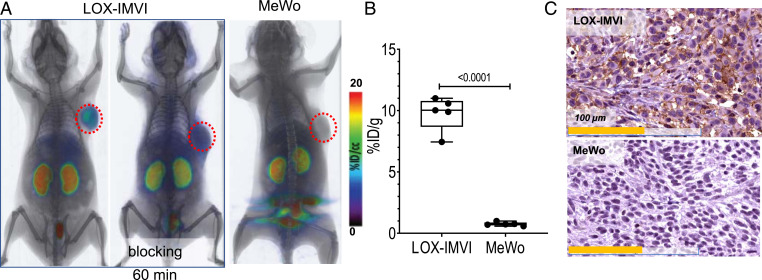
[18F]DK222 PET in mice with human melanoma xenografts shows high-contrast images at 60 min. NSG mice with human melanoma xenografts were injected with ∼200 μCi (7.4 MBq) [18F]DK222 for PET imaging studies or 50 μCi (1.85 MBq) [18F]DK222 for biodistribution studies. Analyses were conducted at 60 min after injection. (A) Whole-body volume rendered PET-CT images show high and specific uptake of [18F]DK222 in LOX-IMVI tumors that express high PD-L1 but not in mice receiving a blocking dose or bearing low–PD-L1–expressing MeWo tumors (n = 3 or 4). (B) Tumor uptake of [18F]DK222 at 60 min, as measured by ex vivo biodistribution studies (n = 5). Error bars indicate SD (n = 4 or 5). Significance was determined by unpaired t test. (C) IHC staining for PD-L1 in LOX-IMVI and MeWo tumors. P values were determined by unpaired t test.

