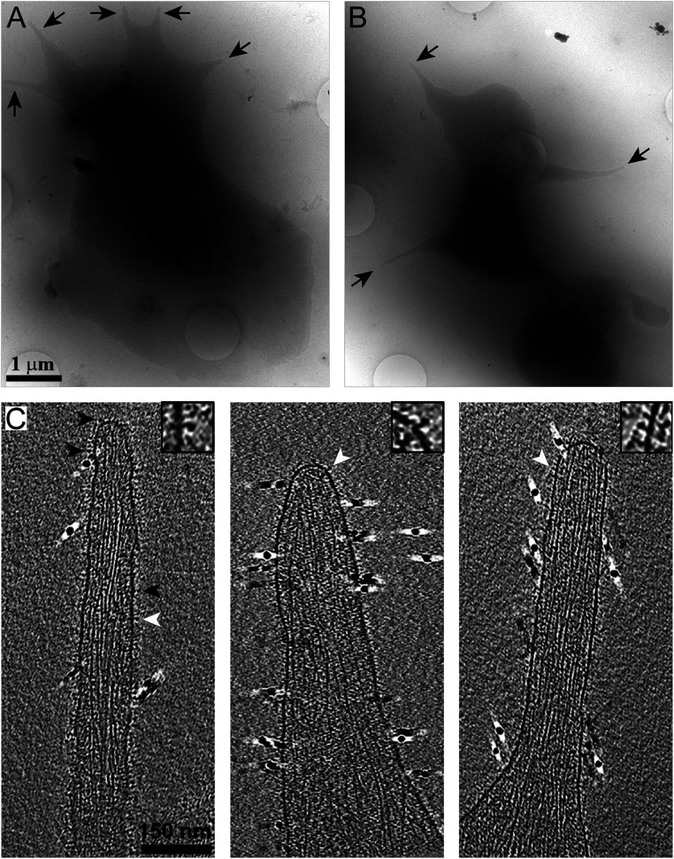
Fig. 1.
Cryo-ET of platelet pseudopodia. (A and B) Cryo-EM of human platelets spread on fibrinogen-functionalized SiO2-coated gold grids. Pseudopodia are detected in single projections (arrows). (C) x-y slices, 10 nm in thickness, through cryotomograms of pseudopodia. The membrane is decorated with protein densities (arrowheads) while the cytoskeleton is seen in the cytoplasm. Insets show magnified membrane receptors (white arrowheads). Fiducial gold clusters are seen as 10-nm black densities.

