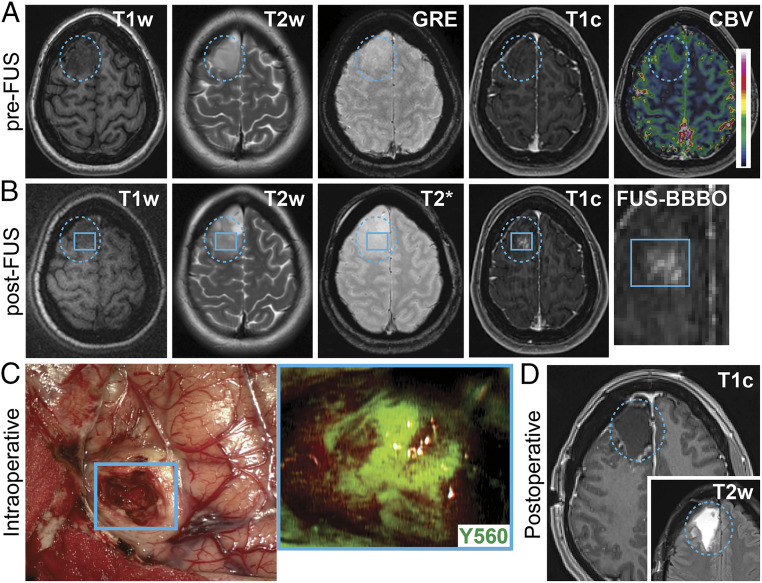
Fig. 2.
Targeted BBBO in infiltrating gliomas: (A) preoperative axial MR images prior to FUS showing T1w, T2w, GRE, T1c, and cerebral blood volume (CBV) sequences. The blue dotted circle indicates the tumor region. (B) Preoperative axial MR images after FUS treatment showing T1w, T2w, GRE/T2*, and T1c sequences. The blue square represents the subspot grid; the inset shows the new contrast enhancement within the target region (e.g., subspot grid). (C) Intraoperative white light images showing the earliest stage of tumor surgery during removal of the MB-FUS–targeted region (blue square). Inset shows fluorescence imaging of this region (visualization score = 3) using the Zeiss YELLOW 560 module. (D) Postoperative axial T1c and T2w MR images showing resection of the intrinsic tumor.

