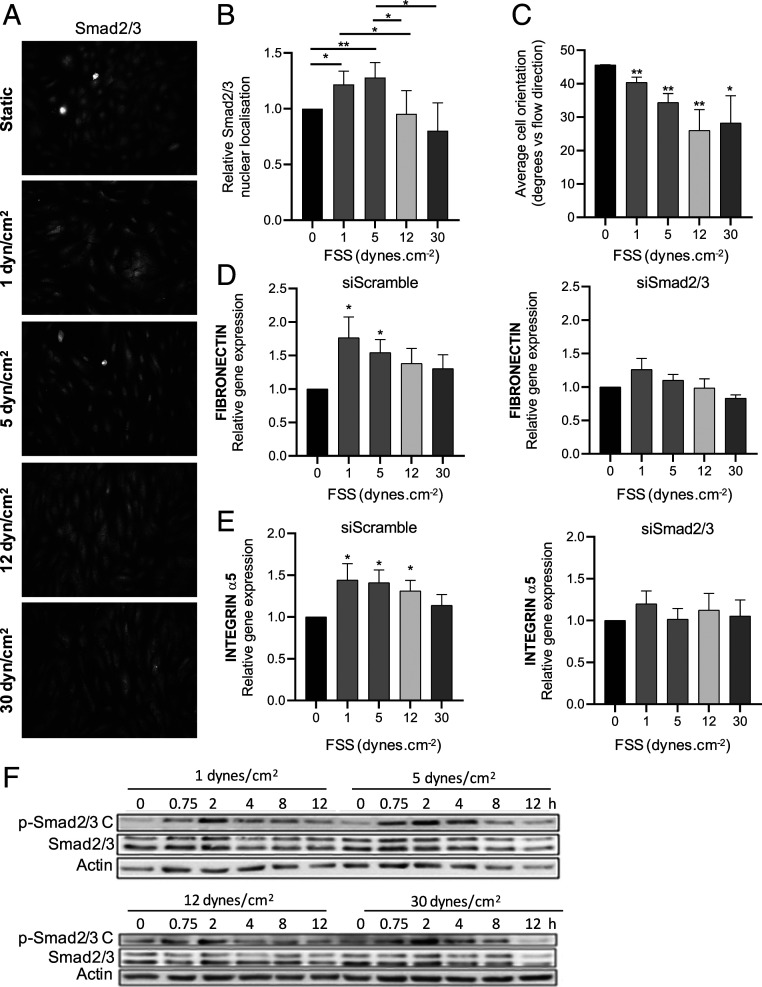
Fig. 1.
Smad2/3 regulation by shear stress. (A) HUVECs were exposed to laminar FSS at the indicated magnitudes for 12 h, fixed, and stained for Smad2/3. (B) Quantification of Smad2/3 nuclear localization, n = 4. (C) Cell alignment was quantified by measuring the angle between the direction of flow and the long axis of the cell; n = 4. (D and E) Fibronectin and integrin α5 mRNA levels were assessed by qPCR; n = 4. (F) HUVECs were exposed to laminar FSS at the indicated magnitudes for 12 h. p-Smad2/3 C was assessed by the Western blotting of cell lysates. Similar results were obtained in three independent experiments. Quantified values are means ± SEM, *P < 0.05, and **P < 0.01, calculated by one-way ANOVA with Tukey’s multiple comparison tests.

