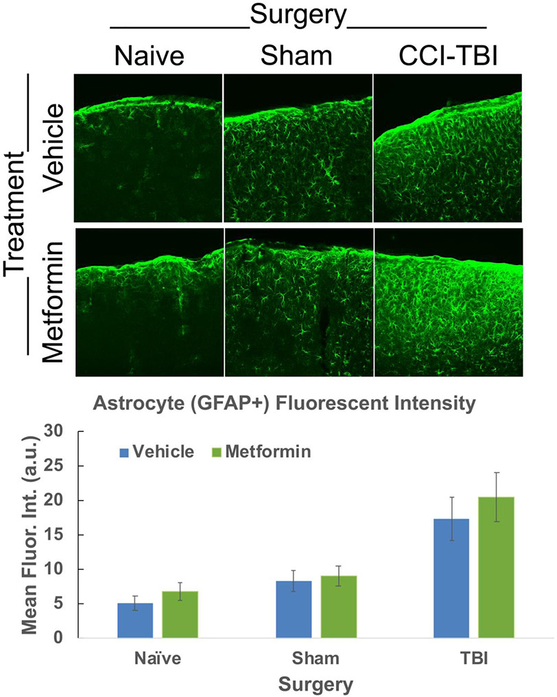
Figure 6. Astrocyte activation is not significantly altered following Metformin treatment.
Confocal images were acquired of the ipsi-lateral cortex of CCI-TBI, sham-operated and naïve control mouse tissue and the fluorescent intensity was manually quantified of GFAP+ cells using ImageJ. No significant difference in fluorescent intensity of GFAP+ labeled astrocytes was found between vehicle or metformin treated Naïve, sham-operated control or CCI-TBI mice. Data represent the mean ± SEM. A 2-way ANOVA was performed. N (animals/images) = Vehicle: Naive=8/24, Sham=12/32, TBI=10/31; Metformin: Naive=10/27, Sham=12/37,TBI=11/36. Data were analyzed by type III test of fixed effects using a linear mixed model analysis. No significant differences were observed between the experimental groups.

