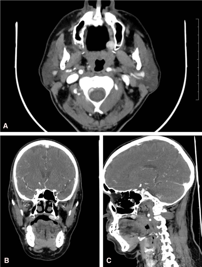
Figure 2.
(A) Axial CT image showing intensely enhancing soft-tissue density swelling in the hard palate at the junction of hard and soft palate on the left. (B) Coronal CT image showing intensely enhancing soft-tissue density swelling in the hard palate at the junction of hard and soft palate on the left. (C) Sagittal CT image showing intensely enhancing soft-tissue density swelling in the hard palate at the junction of hard and soft palate on the left.

