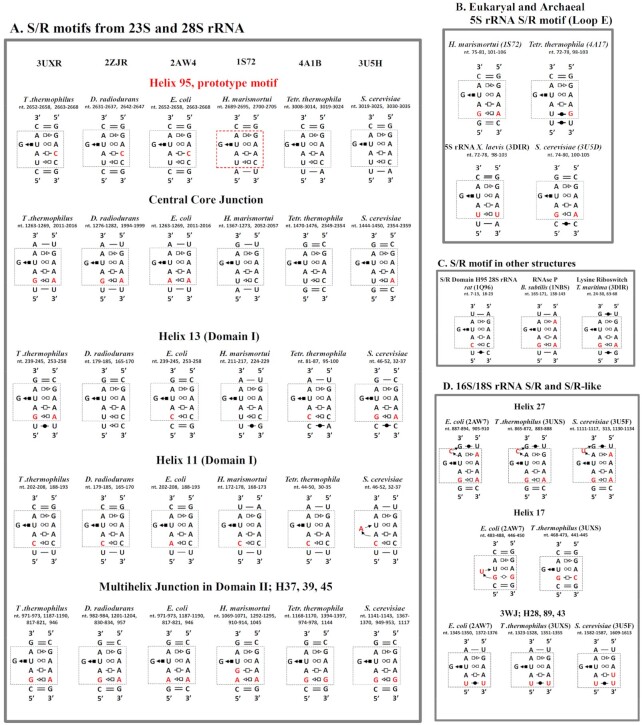
Figure 2.
2D diagrams of S/R motif from different organisms found in 3D rRNA data base. (A) S/R motifs from large ribosomal subunit, the pdb code of the structures is given on the top. Rows represent diagrams of S/R motif corresponding to particular place in the rRNA and name with the nucleotide numbers are given on the top of each diagram. Each column is structure of particular organism. (B) S/R motif diagrams from archaea and eukaryal 5S rRNA, pdb codes are given in parentheses. (C) S/R motif diagram from other structures. (D) S/R-like motifs from 16S rRNA 3D structures of E. coli and T. thermophilus and 18S rRNA of S. cerevisiae.

