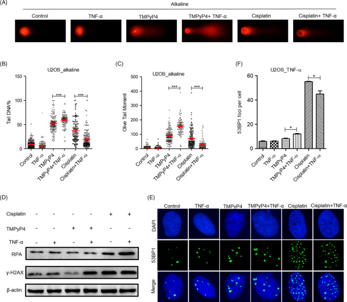
FIGURE 3.

TMPyP4 triggers intense DNA damage and provokes a strong DNA damage response in the inflammatory microenvironment. (A) Representative results of alkaline comet assay. U2OS cells were pre‐treated with 10 μmol L–1 TMPyP4 or 5 μmol L–1 cisplatin for 24 h, 10 ng/mL TNF‐α was added to the wells for 48 h and ≥200 cells were examined in each group. Magnification: 400×. (B) and (C) Quantification of A. (D) Western blot analysis of γ‐H2AX and RPA in U2OS cells exposed to 10 μmol L–1 TMPyP4 or 5 μmol L–1 cisplatin for 24 h and then treated with 10 ng/mL TNF‐α to the appropriate wells for 48 h. β‐actin was used as a control. (E) Immunofluorescence assay was used to determine the 53BPl foci in drug‐treated U2OS cells at the indicated time. Magnification: 400×. (F) Quantification of data in E, ≥200 cells were examined in each group. The values are represented as the mean ± SD of at least three independent experiments. The statistical significance was calculated using the unpaired Student's two‐tailed t test (*p < .05, **p < .01, ***p < .001)
