FIGURE 1.

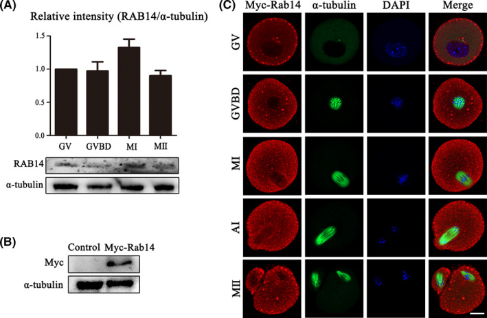
Expression and localization of RAB14 in mouse oocytes. (A) Mouse oocytes at different meiosis stages (GV, MI, ATI and MII) were examined by Western blot. Quantitative analysis of the relative intensity of RAB14 protein expression (relative to that of α‐tubulin) at different stages (GV, GVBD, MI and MII) indicated that RAB14 expressed during oocyte maturation. (B) Protein expression of exogenous RAB14 with an Myc tag was detected in the oocytes after the Myc‐Rab14 mRNA injection. (C) Oocytes from the GV to MII stages were stained with anti‐Myc antibody (red) and counterstained with Hoechest 33342 to visualize DNA (blue). The RAB14 protein was detected in the cytoplasm and cortex during oocyte maturation, and RAB14 was also accumulated at the spindle periphery after GVBD in mouse oocytes. Green, α‐tubulin. Bar = 20 μm
