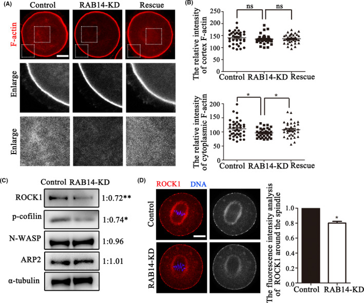
FIGURE 4.

RAB14 knockdown disrupts cytoplasmic actin assembly in mouse oocytes. (A) Representative images of actin filament distribution at the oocyte cortex and cytoplasm in the control group, RAB14‐KD group and rescue group. Red, F‐actin. Bar = 20 μm. (B) No significant difference was observed between control oocytes, Rab14 siRNA‐injected oocytes and rescue oocytes for the F‐actin fluorescent intensities at the cortex (P > .1); however, F‐actin fluorescent intensities in the cytoplasm were decreased in the Rab14 siRNA‐injected oocytes compared with the control and rescue oocytes. *, significant difference (P < .05). (C) Quantitative analysis of the relative intensity of ARP2, N‐WASP, ROCK1 and p‐cofilin in oocytes by Western blot. The results indicated that ROCK1 and p‐cofilin protein expression were significantly reduced in oocytes after RAB14 depletion. **, significant difference (P < .01); *, significant difference (P < .05). (D) Representative images of ROCK1 in the control and RAB14‐KD oocytes. ROCK1 fluorescent intensities decreased in the RAB14‐KD oocytes compared with the control oocytes. *, significant difference (P < .05). Bar = 20 μm
