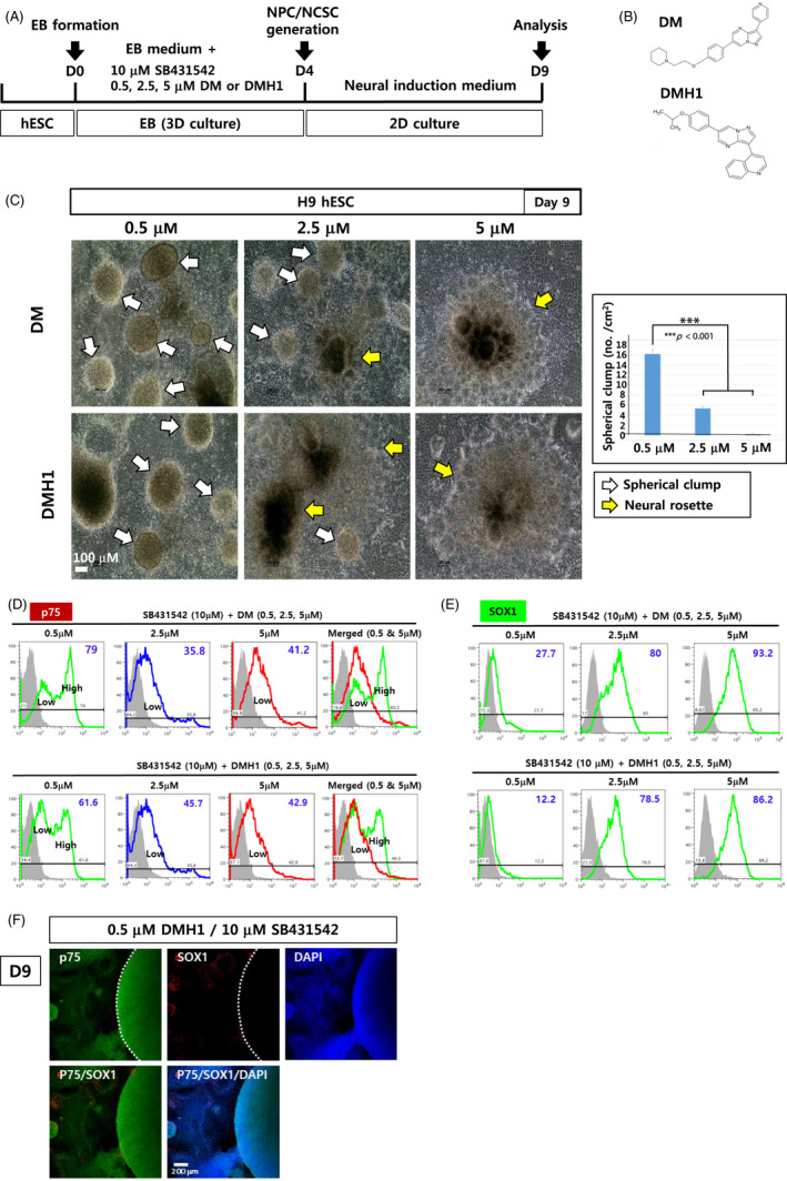
FIGURE 1.

Differentiation of hPSCs into NCSCs using modified dual‐SMAD inhibition. (A) A schematic diagram summarizing the differentiation of H9 hESCs into NPCs and NCSCs. (B) Chemical structures of DM and DMH1. (C) EBs generated in suspension for 4 days in EB medium supplemented with SB431542 (10 μmol/L) and DMH1 (or DM) (0.5, 2.5 and 5 μmol/L) were then attached to dishes for another 5‐d culture. Spherical cell clumps and neural rosettes are marked by white and yellow arrows, respectively. Scale bar: 100 μm. (D, E) After 9 d of differentiation, cells were analysed with flow cytometry using antibodies against p75 (D) and SOX1 (E). (F) Immunostaining of the cells was performed with antibodies against p75 and SOX1 after differentiation with 10 μmol/L SB431542 and 0.5 μmol/L DMH1. Scale bar: 200 μm
