FIGURE 6.

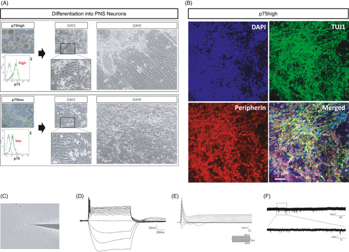
Generation of peripheral neurons from either hESC‐derived p75high or p75low cells. (A) p75high cells (top) and p75low cells (bottom) were obtained from mechanically dissected spherical cell clumps and cell monolayers, respectively. Both cell groups were induced to differentiate into neurons. (B) Neurons derived from p75high cells were immunostained for TUJ1 and peripherin. Scale bar: 50 μm. (C‐F) Electrophysiological analysis of NCSC‐derived peripheral neurons was performed. The morphology of the neurons recorded was shown (C). Representative action potential firing in the peripheral neurons was shown. Scale bar: 20 mV, 200 ms (D). Sodium channel current was shown in the neurons by voltage‐clamp step protocol. Scale bar: 500 pA, 1 ms (E). The trace of spontaneous EPSCs from the neurons was shown. Scale bar: 10 pA, 10 s (upper); 10 pA, 1 s (enlarged trace, bottom) (F)
