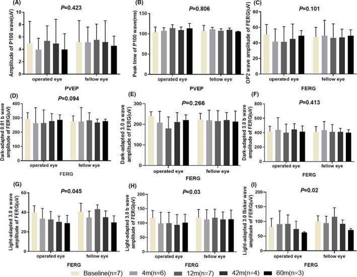
FIGURE 3.

Changes in the visual electrophysiology of the operated and fellow eyes. The amplitude (A) and peak time (B) of P100 wave in the pattern visual evoked potential (PVEP) of the operated and fellow eyes did not significantly differ. Full field electroretinography (ffERG) showed that there was no significant difference between the operated and fellow eyes at same the time point in terms of the amplitudes of Op2 wave (C), b‐wave in dark‐adapted 0.01 (D), a‐wave (E) and b‐wave (F) in dark‐adapted 3.0, a‐wave (G) and b‐wave (H) in light‐adapted 3.0, and wave amplitude of light‐adapted 3.0 flicker (I)
