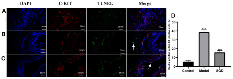
Figure 6.
Results of ICC and TUNEL fluorescence staining in the SO tissue of rabbits (400×, 20 um); Blue is DAPI staining, red is c-kit positive ICC cell staining, green is TUNEL apoptotic cell staining. (A) Normal group, (B) Model group, (C) Treatment group, (D) ICC double positive expression rate. The model group vs the control group, ***P ≤ 0.001. The SGD group vs the model group, ###P ≤ 0.001. Arrows indicate positive expression.

