FIGURE 3.
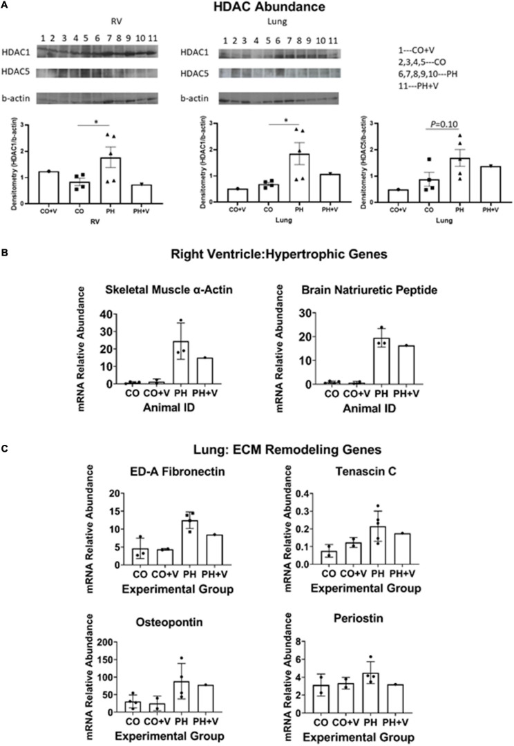
Molecular correlates of vorinostat treatment. (A) Immunoblotting HDAC abundance. RV and lung homogenates were prepared and HDAC abundance was determined by immunoblotting as described in section “Materials and Methods.” Additional untreated control and hypoxic animals were analyzed in parallel for comparison. Protein abundance was quantitated densitometrically relative to beta-actin. *p < 0.05 vs. control (unpaired two-tailed t-test). (B) Representative genes in RV hypertrophic remodeling. Abundances of indicated mRNAs were quantitated by real-time PCR as described in section “Materials and Methods.” Animals used in the present study were analyzed together with samples from additional untreated hypoxic and normoxic calves (n = 4 each) that were managed according to the same animal protocols described in section “Materials and Methods,” and studied around the same period of time. (C) Representative genes in pulmonary remodeling. Determinations were performed as described for (A) and in section “Materials and Methods.”

