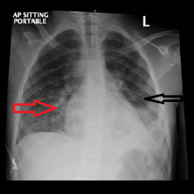
Figure 2.

Chest X-ray 12 h after admission, showing moderatesize patchy area of consolidation in the left lower lobe. Also, there is an area of glass-ground opacity and consolidation noted in the right lower zone medially, suggestive of bronchopneumonia.
