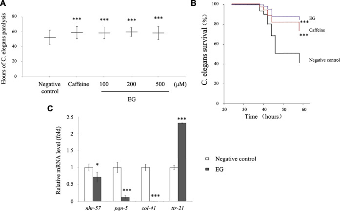
FIGURE 8.
EG retarded transgenic C. elegans paralysis in AD model. (A). CL14176 of C. elegans strain synchronized to L1 phase, and cultured at 16°C for 24 h, then transferred to 25°C to express amyloid peptides that would result in paralysis. The negative control was untreated (equal volume of water), the positive was exposed to 6.27 mM caffeine, and the EG was measured. The mean paralysis times and the ranks of the individual paralysis times in each group are shown (see Supplementary Table S1). Asterisks indicate statistical significance (***p<<0.001). (B) Worms were synchronized and cultured at 16°C for 24 h, then transferred to 25°C for activation. The paralysis phenotypes were detected after 38 h, the number of worms not paralyzed was converted to a percentage, and the “non-paralyzed” percentage was plotted against time activation initiation. The negative control was untreated as described above, the positive was exposed to 6.27 mM caffeine, and the test group to 200 μM EG. Asterisks indicate statistical significance (***p<<0.001). (C) Total RNA of the untreated and EG (200 μM) groups was extracted and reverse transcribed to cDNA. The mRNA levels of nhr-57, pqn-5, col-41, and ttr-21 were measured using quantitative RT-PCR. An asterisk indicates statistical significance (*p < 0.05, **p < 0.001, ***p<<0.001). EG: emodin-8-O-β-d-glucopyranoside.

