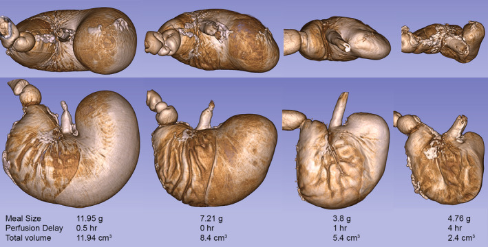
FIGURE 4.

Illustrative micro‐CT images of a variety of iodine stained stomachs (no labeling of the vasculature) at different sizes from full to empty (90 kV; 88 µA; voxel size = 144 µm). The images were created in 3D Slicer from DICOM files. Each stomach is labeled to show the meal size, delay time to perfusion and measured total stomach size. The figure was created by screen capture of the 3D Slicer images followed by import into Photoshop where images were resized with resampling as needed
