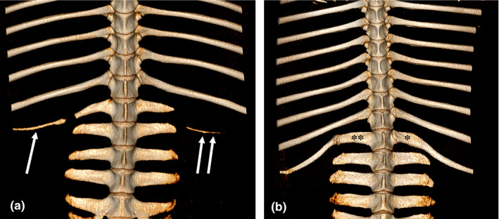
FIGURE 2.

Volume rendering (VR) reconstruction images of the ventral aspect of the caudal thoracic and lumbar vertebrae of two Shetland ponies. (a) (Mare, 4y5m): lumbarization of the last thoracic vertebra. In line with the first right transverse process a remnant of a rib is present (single arrow), on the left side in line with the second transverse process another rib remnant (double arrows) is visible. b) (Mare, 6y): thoracoization of the first lumbar vertebra. Note the difference in location (*,**) of the costovertebral articulations between left and right side
