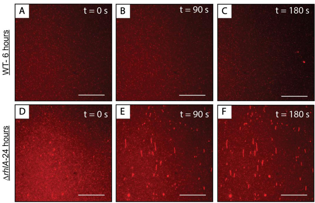
Figure 10:
Representative top-down fluorescence micrographs of structural reformations occurring in fluorescently labeled DOPC SLBs after the introduction of cell-free supernatants of (A-C) wild type P. aeruginosa cultures grown for 6 hours or (D-F) mutant strain ΔrhlA P. aeruginosa cultures grown for 24 hours. The micrographs were acquired at (A, D) 0 seconds, (B, E) 90 seconds, and (C, F) 180 seconds after the first membrane reformations were observed on the surface of the bilayer. The direction of flow in all the images is from the bottom to the top of the screen. Scale bars are 30 μm.

