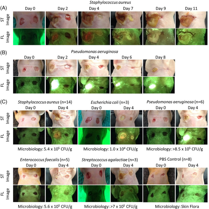
FIGURE 1.

Full‐thickness skin wounds on NCr mice were inoculated with one of five bacterial species (Staphylococcus aureus, Enterococcus faecalis, Pseudomonas aeruginosa, Escherichia coli, Streptococcus agalactiae) or phosphate‐buffered saline as a control. These wounds were imaged with the fluorescence imaging device every 2 to 3 days under standard (ST) and fluorescent (FL) light for up 11 days. In each mouse, the wound on the right was intended to serve as a control, but contamination was observed in many mice. A, Representative images of a mouse inoculated with Staphylococcus aureus. Red fluorescence was observed by Day 2 and peaked at Day 4. B, Representative images of a mouse inoculated with Pseudomonas aeruginosa. Bright cyan fluorescence was observed by Day 1 and remained until the endpoint of the experiment on Day 8. C, Representative standard and fluorescence images of mice inoculated with each bacterial species or PBS negative control at Days 0 and 4 (peak fluorescence). The microbiology analysis shown for each bacterial species was completed at endpoint
