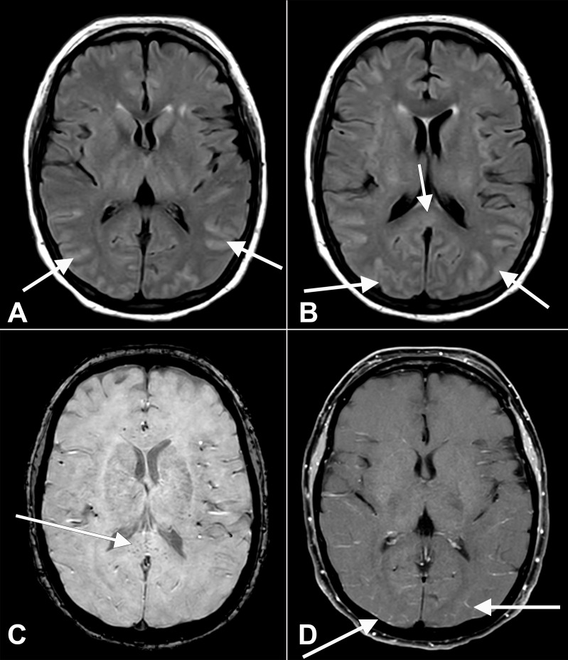
Figure 1.
Axial fluid-attenuated inversion recovery (FLAIR) (A, B) images at the level of the basal ganglia show abnormal FLAIR hyperintense signal (arrows) affecting the bilateral occipital, temporal lobes. This appears almost sulcal suggesting a higher protein component within the cerebrospinal fluid. Note the elevated FLAIR signal in the splenium of the corpus callosum (arrow) suggesting parenchymal insult. Axial susceptibility weighted imaging (SWI) (C) at the level of the splenium of the corpus callosum shows small areas of susceptibility (arrow) in the splenium, likely related to microhaemorrhage. Axial T1 (D) postcontrast with fat suppression at the level of the basal ganglia shows subtle, though true, enhancement (arrows) in the posterior sulci, arachnoid pial (leptomeningeal) pattern suggesting a degree of encephalitis.

