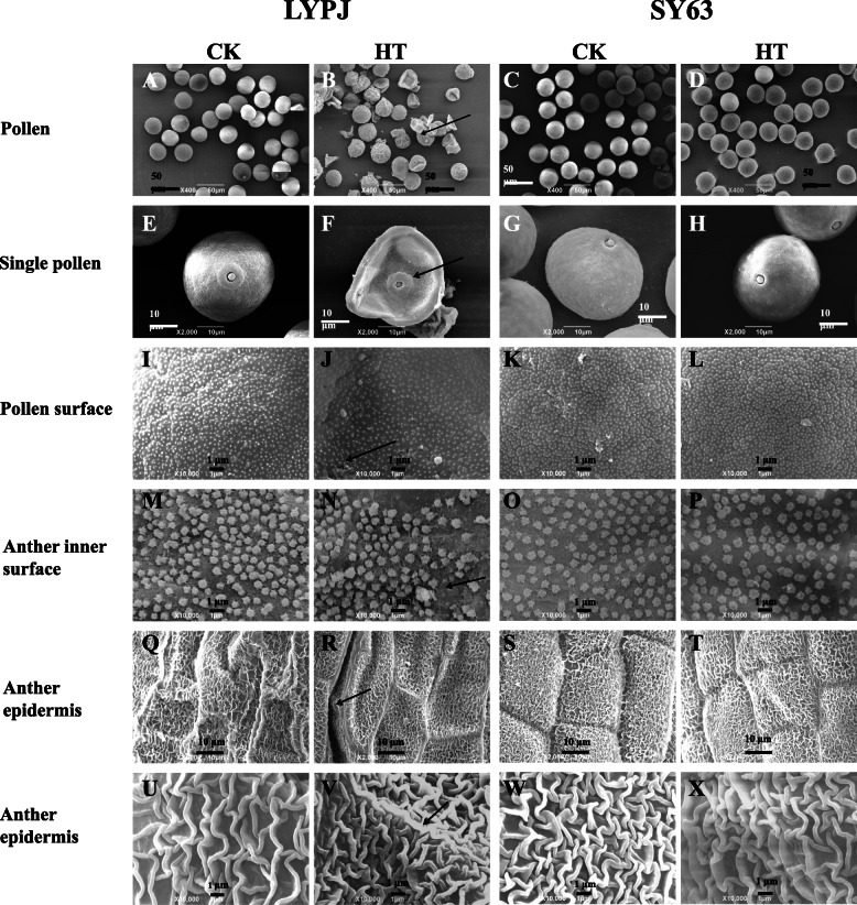
Fig. 2.
Scanning electron micrographs of mature anthers from LYPJ and SY63 plants. A, B, C, and D show scanning electron micrographs of pollen grains (scale bar = 50 μm); E, F, G, and H show magnified single pollen grain (scale bar = 10 μm); I, J, K, and L show the pollen surface (scale bar = 1 μm); M, N, O, and P show the anther inner surface (scale bar = 1 μm); Q, R, S, and T show the anther epidermis at 2000x magnification (scale bar = 10 μm); U, V, W, and X show the anther epidermis at 10000x magnification (scale bar = 1 μm). The arrows indicate shriveled pollen (B), collapsed pollen grain with a hollow germinal aperture (F), abnormal deposition of sporopollenin on the pollen surface (J), uneven distribution of Ubisch bodies on the anther inner surface (N), and compacted anther epidermis (R, V). CK, control temperature; HT, high temperature

