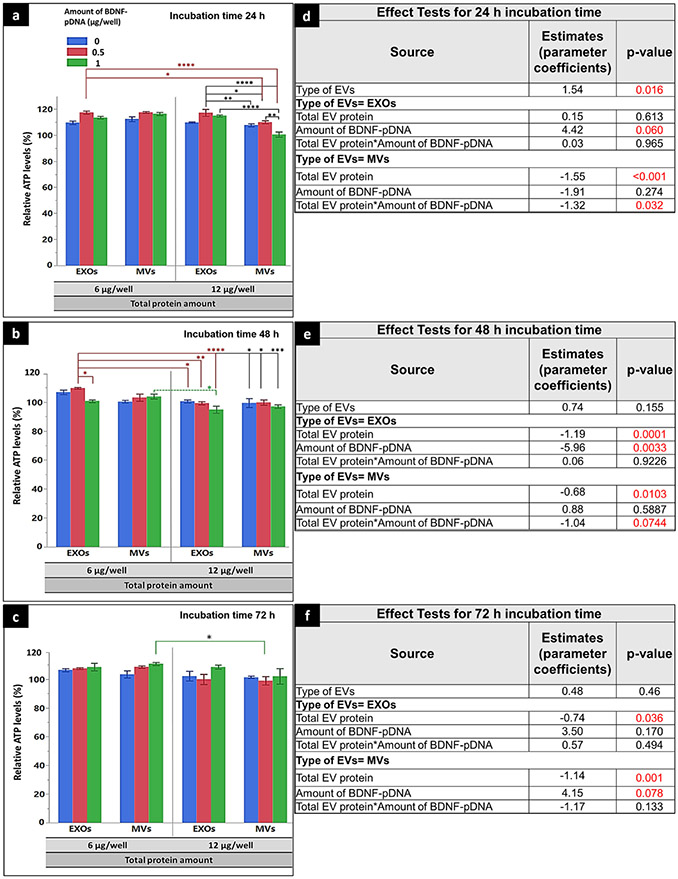
Fig. 10.
Effect of RAW 264.7 cell line–derived naïve EVs or BDNF-EVs on cellular ATP levels in the recipient hCMEC/D3 monolayers. hCMEC/D3 cells seeded at a density of 5000 cell/cm2 in 96 well plates were cultured for 48 h and incubated with BDNF-EXOs, or MVs derived from RAW 264.7 cells pre-transfected with 0 (naïve), 0.5, or 1 μg BDNF-DNA/well. EXOs or MVs were incubated for 24 h (a and d), 48 h (b and e), or 72 h (c and f) at either 6 μg or 12 μg EV protein/well. Post-incubation, the relative ATP levels were measured using a CellTiter-Glo 2.0-based ATP assay. Relative light units (RLU) of the cell lysates were measured at 1 s integration time using a Synergy HTX multi-mode reader. The average relative ATP levels compared with control, untreated cells from the recipient cells treated with DNA-EVs isolated from donor cells transfected with different amounts of BDNF pDNA (0, 0.5, or 1 μg/well) at different doses (6 or 12 μg EV protein/well) when incubated for 24 (a, d), 48 h (b, e), and 72 h (c, f). The effect test using multiple regression analysis presenting parameter estimates (coefficient), and p values of each transfection factor and their two-way interactions for 24-h (d), 48-h (e), and 72-h incubation time (f). Data are presented as mean ± SE of three independent experiments (n = 3/experiment). Statistical comparisons were made using multiple regression analyses, where *p < 0.05, **p < 0.01, ***p < 0.001, and ****p < 0.0001

