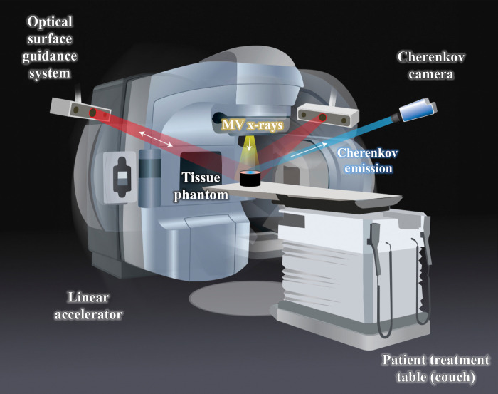
Fig. 6.
Illustration of experimental setup. The linear accelerator irradiates a tissue phantom placed on the couch with MV x-rays, and the gantry can rotate around the couch. The Cherenkov camera fixed to the ceiling captures the Cherenkov emission from the phantom. The clinical setup also includes an optical surface guidance system that is not utilized during these measurements.

