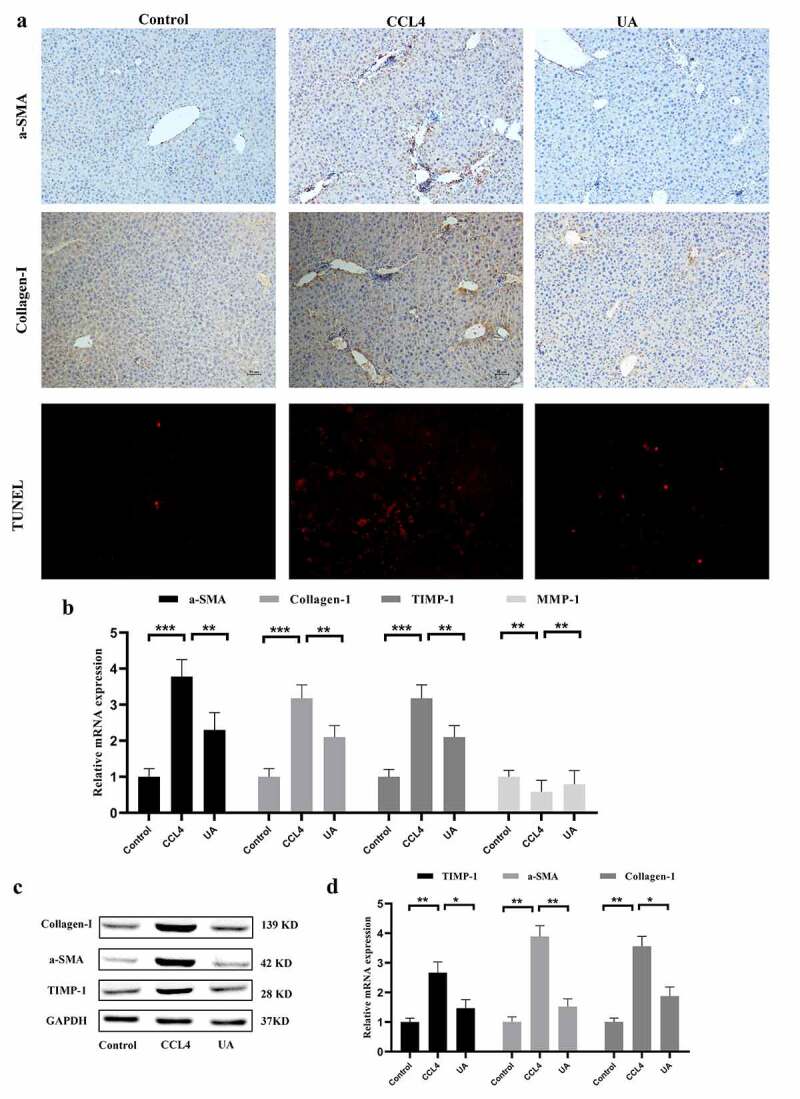
Figure 2.

UA exerts antifibrotic effects by impacting the expression of fibrosis-related factors. (a): IHC was used to measure the levels of α-SMA and Collagen-1. Hepatocyte apoptosis was assessed by TUNEL assays; (b): The expression of α-SMA, Collagen-1, TIMP-1 and MMP-1 was determined by qRT-PCR; (c,d): The levels of Collagen-1, TIMP-1 and MMP-1 were determined by Western blotting. n = 6 per group. The data are presented as the means±SD in each group. *P < .050, **P < .010 and ***P < .001
