Figure 1.

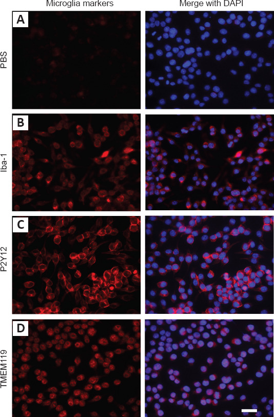
IMG cells were identified by different microglia markers.
(A) PBS (negative control). (B) Iba-1 (Alexa Fluor-568, red). (C) P2Y12 (Alexa Fluor-568, red). (D) TMEM119 (Alexa Fluor-568, red). DAPI, blue. The microglia markers, labeled with red fluorescence, were clearly expressed in the cytoplasm compared with the negative control. Scale bar: 20 μm. DAPI: 4′,6-Diamidino-2-phenylindole; Iba-1: ionized calcium-binding adapter molecule 1; P2Y12: P2Y metabotropic G-protein-coupled purinergic receptors 12; PBS: phosphate-buffered saline; TMEM119: transmembrane protein 119.
