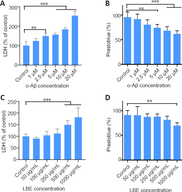
Figure 2.

LDH and PrestoBlue assay responses to o-Aβ or LBE treatment in IMG cells.
(A–D) IMG cells were treated with different concentrations of o-Aβ (A, B) or LBE (C, D) for 24 hours. Cytotoxicity was determined from the culture supernatants using an LDH assay kit. (B, D) Cell viability was evaluated using the PrestoBlue assay and are represented as the percentage of survival. Data are expressed as the mean ± SD from four independent experiments. **P < 0.01,***P < 0.001 (one-way analysis of variance followed by Tukey’s post hoc test). Aβ: Amyloid-β; IMG: immortalized microglial cell line; LBE: Lycium barbarum extract; LDH: lactate dehydrogenase; o-Aβ: oligomeric Aβ1–42.
