Figure 4.
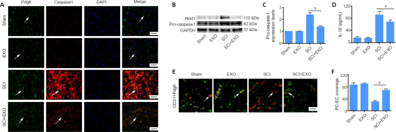
BMSC-EXOs decrease pyroptosis in pericytes and increase pericyte coverage in vivo.
(A) Colocalization of Pdgfr and caspase 1 in the spinal cord at 3 dpi. Arrows indicate pericytes. Red: DyLight 594, caspase 1; green: DyLight 488, Pdgfr; blue: DAPI. (B, C) Expression of Nod1 (data not shown) and pro-caspase 1 (fold-change compared with the Sham group) assessed by western blot assay. (D) Expression of IL-1β assessed by enzyme-linked immunosorbent assay. (E) Representative immunofluorescence images of Pdgfrβ+ pericytes and CD31+ endothelial cells after SCI. Compared with the Sham and EXO groups, the SCI group showed loss of pericytes and low PC/EC coverage. Exosome treatment reduced these changes. Arrows indicate microvessels. Red: DyLight 594, CD31+ endothelial cells; green: DyLight 488, Pdgfrβ+ pericytes; blue: DAPI. Nuclei were labeled with DAPI. Scale bars: 50 µm in A, and 20 µm in E. (F) Percentage of PC/EC coverage in spinal cord microvessels. Data are expressed as the mean ± SD (n = 3 in C, n = 6 in D and F). *P < 0.05 (Student’s t-test). BMSC-EXO: Bone mesenchymal stem cell-derived exosome; DAPI: 4′,6-diamidino-2-phenylindole; EXO: exosome; PC/EC: pericyte/endothelial cell; Pdgfr: platelet-derived growth factor receptor; SCI: spinal cord injury.

