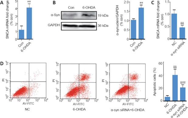
Figure 2.

6-OHDA exerts neurotoxicity by upregulating α-synuclein expression.
(A) Reverse transcription quantitative polymerase chain reaction analysis of SNCA mRNA levels in SH-SY5Y cells after treatment with 6-OHDA for 24 hours; GAPDH served as the internal control. (B) Representative western blot and quantification data of α-synuclein in SH-SY5Y cells treated with 6-OHDA for 24 hours. (C) Reverse transcription quantitative polymerase chain reaction analysis of SNCA mRNA levels in SH-SY5Y cells transfected with SNCA siRNA for 24 hours. (D) Cell apoptosis was assessed using Annexin V-FITC/propidium iodide. The data are shown as the mean ± SD (n = 5; A–C: Student’s t-test; D: one-way analysis of variance followed by Bonferroni post hoc test); **P < 0.01, vs. control group; ##P < 0.01, vs. 6-OHDA treatment group; &P < 0.05; &&P < 0.01, vs. NC group. 6-OHDA: 6-Hydroxydopamine; Con: control; GAPDH: glyceraldehyde 3-phosphate dehydrogenase; NC: negative control; siRNA: small interfering RNA; SNCA: the gene encoding α-synuclein; TGIF1: TG-interacting factor 1; α-syn: α-synuclein.
