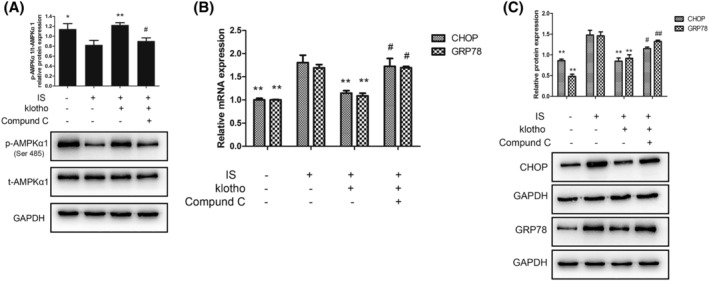
FIGURE 3.

(A) Effects of IS and Klotho on the phosphorylation of AMPKα1. Cells were pretreated with Klotho (100 μg/L) for 1 h, then incubated with IS (50 μg/mL) for further 48 h with or without Compound C (10 μmol/L). (A) Western blotting analysis for p‐AMPKα1 and t‐AMPKα1. Data were shown as mean ± SD. Statistical differences were expressed as * P < 0.05 versus IS alone; ** P < 0.01 versus IS alone; #P < 0.05 versus IS and Klotho treatment together; ##P < 0.01 versus IS and Klotho treatment together. (B,C) Effects of IS and Klotho on the expression of GRP78 and CHOP in HUVECs. Cells were pretreated with Klotho (100 μg/L) for 1 h, then incubated with IS (50 μg/mL) for further 48 h with or without Compound C (10 μmol/L). (B) Quantitative polymerase chain reaction (qPCR) analysis for GRP78 and CHOP. (C) Western blotting analysis for GRP78 and CHOP. Data were shown as mean ± SD. Statistical differences were expressed as *P < 0.05 versus IS alone; **P < 0.01 versus IS alone; #P < 0.05 versus IS and Klotho treatment together; ##P < 0.01 versus IS and Klotho treatment together. HUVECs, human vein umbilical endothelial cells; IS, indoxyl sulfate; t‐AMPKα1, total AMP‐activated protein kinase α1; p‐AMPKα1, phosphorylated AMP‐activated protein kinase α1; GRP78, glucose‐regulated protein 78; CHOP, CCAAT/enhancer binding protein homologous protein
