
Figure 1.
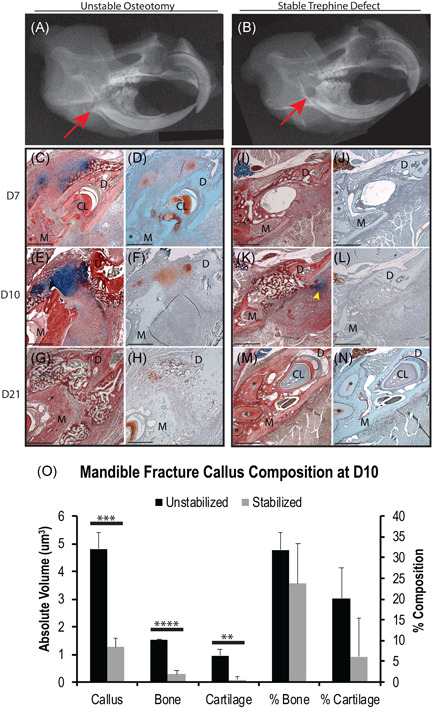
Fracture stability mediates mandibular ossification mechanisms. X‐ray images of an unstable transverse osteotomy (A) and stable 1 mm trephine defect (B) confirm the proper anatomical location of fracture site (red arrow) within the right mandibular ramus of C57BL/6 mice. Mandibles were imaged at 7 days postfracture (N = 5/group). HBQ (C, E, G, I, K, M) and Saf‐O (D, F, H, J, L, N) standard histology staining of transverse sections reveals that mandibles with unstable osteotomies (C–H) develop a robust cartilage callus and primarily heal through endochondral ossification, whereas mandibles with stable trephine defects (I–N) develop little to no cartilage and primarily heal through intramembranous ossification. Small amounts of cartilage were found adjacent to the fracture site in the stabilized fracture model at 10 days postfracture, usually ventral and posterior to the defect (K, yellow arrowhead). Cartilage was only observed in the stable fracture model at day 10 postfracture and was most‐likely due to motion derived from residual bone chips left behind from drilling the trephine defect. Stereological analysis of callus tissue composition confirmed that the absolute volume of cartilage was significantly less in stable than in unstable fractures at D10, which is the time point during which the greatest amount of cartilage was observed in either model (O). Complete bony‐bridging was observed in the unstable model at D21 postfracture (G, H) and in the stable model at D10 postfracture (K, L). HBQ: cartilage is blue, bone is red. Saf‐O: cartilage is red, bone is teal. (*) in histology = mandibular molar roots. N = 5/time point/fracture type. Scale = 500 µm. **p < .01. ***p < .001. ****p < .0001. CL, cervical loop; D, distal; HBQ, hall brunt quadruple; M, mesial; Saf‐O, Safranin‐O [Color figure can be viewed at wileyonlinelibrary.com]