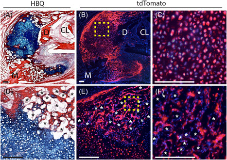
Figure 2.

Fracture callus chondrocytes give rise to osteoblasts and bone‐lining cells during mandibular endochondral repair. Lineage tracing of mandibular fracture callus chondrocytes was performed using the Aggrecan‐CreERT2driver to induce cartilage‐specific Cre recombination in the stop‐floxed Ai9 tdTomato reporter mouse. Cre recombination was induced with daily Tamoxifen injections from days 6‐10 postfracture with samples harvested at 14 days postfracture. Fluorescence microscopy indicates robust tdTomato expression in chondrocytes within the fracture callus indicating highly efficient Cre recombination (C, boxed region in B). Robust tdTomato expression is also observed in osteoblasts/cytes within the newly formed bone and in bone‐lining cells, confirming their chondrocyte‐derivation (F, boxed region in E). Low‐magnification images demonstrate the significant contribution of chondrocytes to new‐bone formation (A, D correspond to B, E). DAPI counterstain was used to visualize nuclei (blue). *Blood vessels. N = 5. Scale = 100 µm. CL, cervical loop; D, distal; DAPI, 4′,6‐diamidino‐2‐phenylindole; M, mesial [Color figure can be viewed at wileyonlinelibrary.com]
