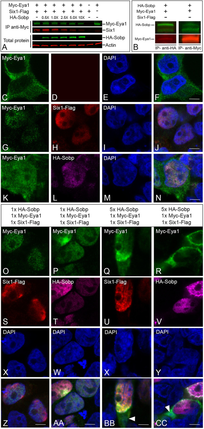
Fig. 2.

Sobp reduces the transcriptional activation of Six1+Eya1 target genes by disrupting the Six1/Eya1 interaction. (A) HEK293T cells co-transfected with equimolar amounts of Six1-Flag and/or Myc-Eya1 were additionally transfected with increasing amounts of HA-Sobp. Although low (0.5×) and equimolar (1.0×) levels of Sobp resulted in higher levels of Six1 bound to Eya1 relative to when Six1 and Eya1 were co-transfected without Sobp, the amount of Six1 bound to Myc-Eya1 decreased with increasing levels of HA-Sobp (2.5×, 5.0× and 10×). The bottom two rows show expression before immunoprecipitation of increasing levels of HA-Sobp with β-actin as loading control. (B) HEK293T cells were co-transfected with HA-Sobp and Myc-Eya1 followed by multiplex fluorescence western blot detection for HA-Sobp (green) and Myc-Eya1 (red). Myc-Eya1 is detected when HA-Sobp is immunoprecipitated (IP, anti-HA, left column). The reverse immunoprecipitation (anti-Myc, right column) confirmed this interaction. (C-N) Confocal images of HEK293T cells expressing Myc-Eya1 (green), Six1-Flag (red) and/or HA-Sobp (magenta). Myc-Eya1 is located exclusively in the cytosol (C,F) and is completely translocated to the cell nucleus by Six1-Flag in the majority of the cells (G,H,J). Surprisingly, HA-Sobp also partially translocates Myc-Eya1 to the cell nucleus (K,L,N) in the absence of Six1. Cell nuclei are stained with DAPI (blue in E,F,I,J,M,N). Scale bars: 5 μm. (O-CC) Confocal images of HEK293T cells expressing Myc-Eya1 (green, O-R,Z-CC), Six1-Flag (red, S,U,Z,BB) and HA-Sobp (magenta, T,V,AA,CC). Myc-Eya1 was completely translocated into the cell nucleus by Six1 when cells received equimolar amounts (1×) of Six1-Flag, Myc-Eya1 and HA-Sobp (O,P,S,T,Z,AA), whereas cytosolic Myc-Eya1 (arrowheads in BB and CC) was detected in many cells when there was a fivefold increase in HA-Sobp. Nuclear DAPI staining, blue (X-CC). Scale bars: 5 μm.
