
Fig 2. Scanning electron microscopy (SEM) of leptospiral renal biofilm and its matrix in wild naturally colonized Rattus norvegicus.
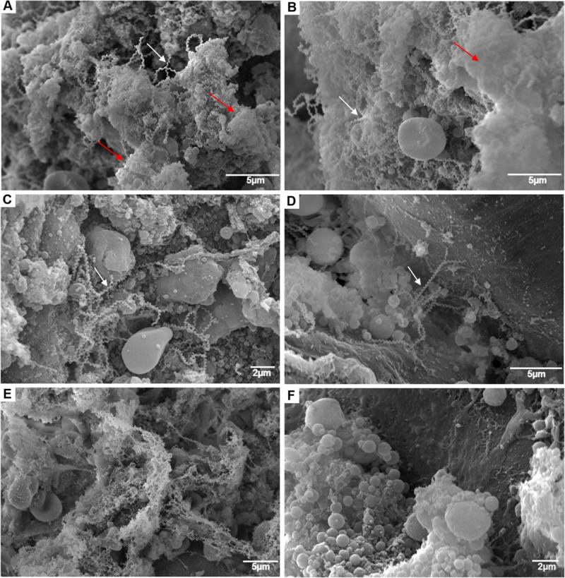
A and B–SEM of colonized kidney with ruthenium red (RR) showed Leptospira agglomerates (white arrows) surrounded by an anionic exopolysaccharidic matrix (red arrows) inside the renal tubules. C and D–SEM of colonized kidney without RR, where leptospires are evidenced agglomerated (C) or isolated (D), without the presence of the matrix. E–SEM using RR of R. norvegicus negative control. F–SEM without RR of R. norvegicus negative control.