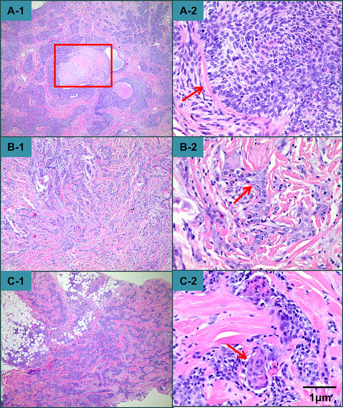
Figure 3.
Histopathological examination revealed that the nodule exhibited epithelial tumor islands interspersed with a dense fibrous connective tissue stroma and centrally located trichilemmal keratinization. (A-1) At higher magnification, the tumor had polygonal cells, palisade arrangements with focal inversion, a thick hyaline membrane surrounding each lobule, and atypical cytology. A high mitotic index and atypical mitotic figures were observed. (A-2) The presentation of the plaque and the mass on the neck was distinct from that of the nodular lesion on the scalp. There were some cable-like structures, which may have even reached from the dermis to the subcutaneous fat in the mass on the neck (B-1, B-2, C-1, C-2).

