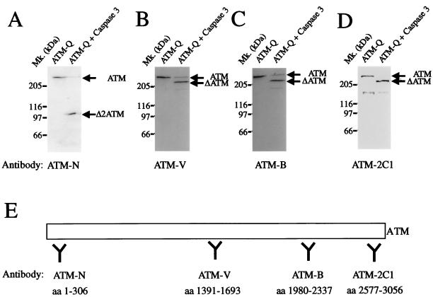
FIG. 5.
Antibody mapping of ATM fragments generated by caspase 3. Partially purified ATM (ATM-Q; 50 μg of total protein) was incubated with 300 ng of recombinant caspase 3 for 30 min at 37°C. Western blot analysis was performed with polyclonal rabbit antiserum raised against the N-terminal portion of ATM (ATM-N) (A), the central region of ATM (ATM-V) (B), the C-terminal region of ATM (ATM-B) (C), or the kinase domain of ATM (ATM-2C1) (D). (E) Schematic representation of ATM and the regions against which the antibodies were raised. Mk., molecular size markers; aa, amino acids.

