
FIGURE 1.
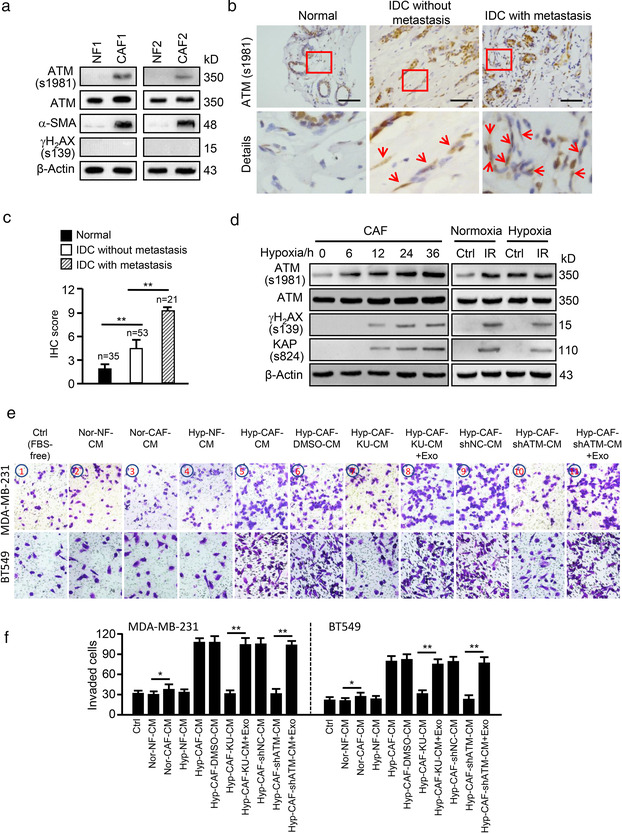
Hypoxia‐mediated oxidized ATM in breast CAFs promotes breast cancer cell invasion. (a) Protein levels of ATM, ATM (s1981), α‐SAM and γH2AX (s139) were detected by western blotting in paired primary NFs and CAFs from two patients. (b) Representative images of activated ATM (oxidized ATM) proteins in stromal fibroblasts of clinical breast tumour tissues (Scale bar, 200 μm; Red arrow, oxidized ATM). (c) Quantification of oxidized ATM in stromal fibroblasts of BC tissues. (d) Western blotting to determine activated ATM, total ATM, γH2AX (s139) and KAP (s824) protein levels in CAFs under hypoxia treatment for the indicated hours or in CAFs exposed to 0.5 Gy ray irradiation (IR) as a control group. (e) MDA‐MB‐231 and BT549 cells were co‐cultured with the CM from normoxic or hypoxic NFs and CAFs; or CM from hypoxic CAFs treated with or without KU60019 (KU:KU60019), or CM from ATM wild type (shNC) or silenced (shATM) hypoxic CAFs. Exosomes isolated from hypoxic CAF (Hyp‐CAF‐Exo: Exo) were added to the co‐culture system, and cell invasion potentials were evaluated by Transwell assay. Ctrl: Tumour cell + FBS‐free medium. (f) Quantification of invaded cells in (e) (*P < 0.05; **P < 0.01.)