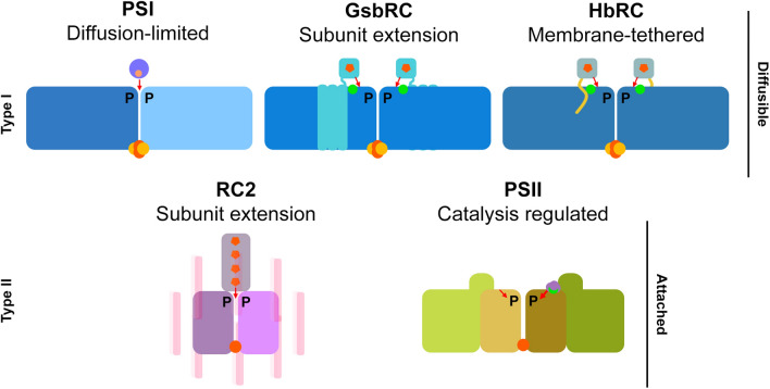
Fig. 2.
Themes of electron donation in RCs. Modes of electron donation and directionality (red arrows) are shown. Type I RCs (top row) exhibit only diffusible electron donors and type II RCs exhibit bound electron donors. Ca2+ sites are shown with green spots. Hemes are shown as orange pentagons. Core-associated [4Fe-4S] clusters are show as orange and yellow spheres. Non-heme irons are shown as single orange spheres. A tetraheme cytochrome is commonly found in anoxygenic RC2s, however, cases in which this subunit has been lost are known within the Proteobacteria

