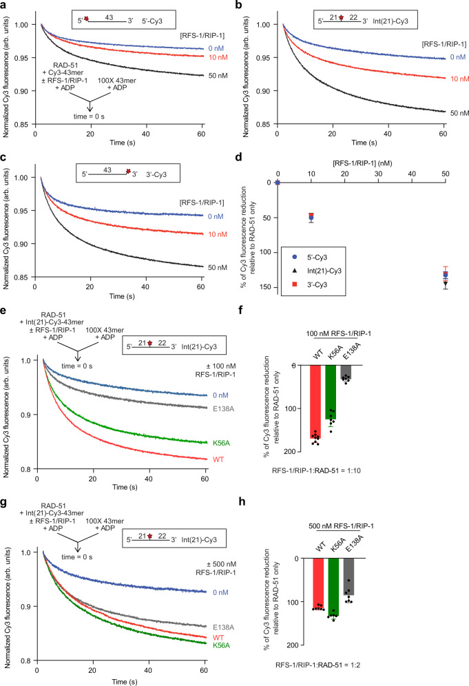
Fig. 4. RAD-51 destabilization by RFS-1/RIP-1 occurs throughout the whole filament and is partially dependant on the RFS-1 Walker boxes.
a–c Average normalized Cy3-43mer fluorescence profiles plotted as a function of time. The arrow indicates the components of the two syringes rapidly mixed at the 0 s time point in a stopped flow instrument. Schematics of the different Cy3 label positions are shown inset. RAD-51-ssDNA filaments pre-formed with RAD-51 (1 μM), Cy3-43mer ssDNA (15 nM) and indicated concentrations of RFS-1/RIP-1 in the presence of ADP for 10 min were mixed with 100-fold excess unlabelled 43mer. (a) 5′-, (b) Int(21) or (c) 3′-end Cy3 labelled substrate was used (a: n = 17–20, b: n = 14–16, c: n = 15–16). d Graph of RFS-1/RIP-1 concentration-dependence of Δ Cy3 fluorescence for the data presented in (a–c) (mean; errors: s.d.). e–h Comparison of WT and Walker box mutants of RFS-1 (E138A and K56A) using two different concentrations of RFS-1/RIP-1: 100 nM (e) (n = 7–9) and 500 nM (g) (n = 4–7). Experiments were done similarly as in a–c. f, h Quantification of the data over the full-time course of the experiments in (e and g) respectively (mean; errors: s.d.). Source data are provided as a Source Data file.

