Figure 1.
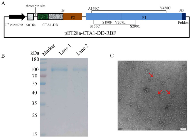
Design and analysis of the CTA1-DD-RBF protein. (A) Domain structure of the CTA1-DD-RBF protein. Mutations (Val to Leu at residue 207 and Ser to Phe mutation at residue 190; disulfide between residues 290 and 155) and disulfide bonds 149 to 458 were incorporated into the F protein and are indicated by vertical black lines. CTA1-DD-RBF contains the CTA1 subunit of cholera toxin (CT), two immunoglobulin (Ig) binding domains (DD) of staphylococcal protein A and hRSV F protein (residues 26–105 and 146–513) with a T4 fibritin trimerization motif (foldon) and a variable linker, GSGSG. (B) CTA1-DD-RBF proteins digested by thrombin on SDS-PAGE: Marker: protein markers; Lane 1: CTA1-DD-RBF purified using a HisTrap FF column after digested by thrombin; Lane 2, CTA1-DD-RBF purified by HisTrap FF columns before digested by thrombin. (C) Negative stain electron microscopy of CTA1-DD-RBF. The CTA1-DD-RBF was highly homogenous. Bar = 50 nm.

