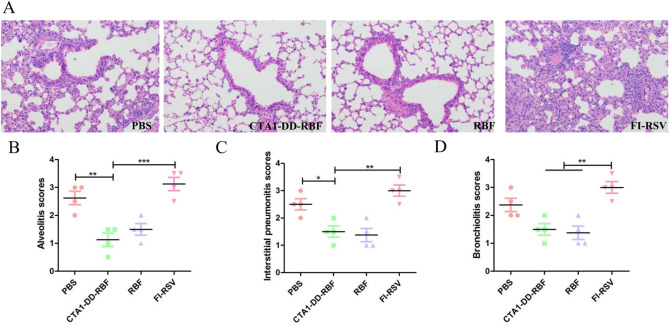
Figure 6.
Histopathological analysis of hematoxylin and eosin (H&E)-stained lungs. The left lungs were dyed with H&E for histological assessment. All images were obtained at 200 × magnification. (A) The left lungs were dyed with H&E for histological assessment. (B) Scoring of alveolitis in immunized mice after hRSV challenge. (C) Scoring of interstitial pneumonitis in immunized mice after hRSV challenge. (D) Scoring of bronchiolitis in immunized mice after hRSV challenge. The degree of inflammation in the alveolar tissue was graded as follows: 0, normal; 1, mild inflammation; 2, moderate inflammation; 3, marked inflammation; and 4, severe inflammation. Statistically significant differences were determined by one-way ANOVA with the Newman-Keuls posttest. *p < 0.05, **p < 0.01, ***p < 0.001. The bars represent the mean values with standard deviations.

